
Why early detection matters before cirrhosis develops
Liver fibrosis: what happens to the tissue of the organ
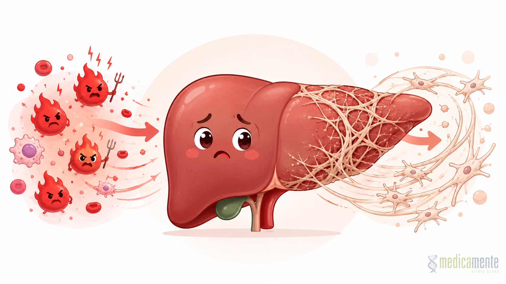
Liver fibrosis is not a separate disease, but a universal response of the liver to long-term damage. When liver cells face chronic inflammation, viral infection, metabolic overload, toxic exposure or autoimmune attack, the body attempts to repair the damaged tissue. In the early stages, this process may be protective. But if the damaging factor persists for a long time, repair becomes abnormal: instead of normal liver architecture, connective tissue gradually accumulates.
Connective tissue itself does not perform the functions of hepatocytes. It does not fully participate in detoxification, protein synthesis, fat and carbohydrate metabolism, bile production or storage of energy resources. The more connective tissue appears in the liver, the more strongly its structure is disrupted. This is why fibrosis is considered an important intermediate stage between chronic liver disease and cirrhosis.
It is important to understand that fibrosis does not appear instantly. It is a long process that develops over months and years. In one patient, it may progress slowly; in another, much faster. The rate depends on the cause of liver damage, inflammatory activity, age, genetic factors, alcohol use, the presence of diabetes mellitus, obesity, viral coinfections and other associated conditions.
Why fibrosis can remain unnoticed for a long time
One of the main features of the liver is its high capacity for compensation. Even if part of the tissue has already been damaged, the remaining cells can continue to perform the main functions for a long time. Therefore, a person may feel well, maintain normal daily activity and not suspect that fibrosis is forming. The absence of pain or pronounced weakness does not mean that the liver is completely healthy.
In the early stages, fibrosis usually has no specific symptoms. Sometimes fatigue, discomfort in the right upper abdomen, reduced exercise tolerance or nonspecific changes in blood tests may occur. But such signs do not reliably show whether fibrosis is present or how advanced it is. Therefore, diagnosis should not be based only on how the patient feels.
The danger of fibrosis lies in its hidden progression. While the patient does not feel significant problems, the liver tissue may gradually change, microcirculation may worsen and the inflammatory process may intensify. If this stage is missed, the disease may progress to cirrhosis, where the possibilities of reverse recovery become much more limited.
Main causes of liver fibrosis
Fibrosis can develop in different liver diseases. Its cause must always be identified because treatment is directed not at scar tissue itself, but at the factor that maintains damage.
The most common causes include:
- Chronic viral hepatitis.
Hepatitis B, C and D can maintain inflammation in the liver for years. With active viral replication and a long-term inflammatory process, the risk of fibrosis increases. - Fatty liver disease.
Metabolic dysfunction-associated fatty liver disease is linked to insulin resistance, obesity, type 2 diabetes and lipid metabolism disorders. When steatosis progresses to steatohepatitis, inflammation and fibrosis formation begin. - Alcohol-related liver injury.
Regular toxic exposure to alcohol can cause inflammation, fatty transformation of the liver, fibrosis and cirrhosis. The risk depends on dose, duration of use, individual sensitivity and associated factors. - Autoimmune liver diseases.
In autoimmune hepatitis, primary biliary cholangitis and primary sclerosing cholangitis, damage is related to an abnormal immune reaction or injury to the bile ducts. - Drug-induced and toxic injury.
Some medications, toxic substances and long-term damaging exposures may lead to chronic inflammation and fibrosis. - Hereditary and metabolic diseases.
These include hemochromatosis, Wilson disease and other metabolic disorders in which substances that damage liver tissue accumulate in the liver.
Sometimes several causes are present in one patient at the same time. For example, chronic hepatitis B may coexist with fatty liver disease, and metabolic risk may coexist with regular alcohol use. In such situations, fibrosis may progress faster, so it is important to assess not one cause, but the whole clinical picture.
How inflammation turns into scar tissue
The formation of fibrosis is connected with the activation of special liver cells involved in tissue repair. In short-term damage, they help organize healing. But in chronic inflammation, these cells begin actively producing components of the extracellular matrix, especially collagen. Gradually, connective tissue accumulates where normal liver structure should be preserved.
This process can be understood as abnormal healing. If the skin is damaged once, a small scar forms. If damage repeats constantly, scar tissue becomes more pronounced and changes the structure of the area. A similar process occurs inside the liver. Connective tissue disrupts normal connections between cells, compresses vascular structures and changes blood flow through the liver.
In the early stages, fibrosis may be limited. But as it progresses, connective tissue forms septa that divide liver tissue into areas. When regenerative nodules and gross architectural distortion appear, the condition is already called cirrhosis. This is why fibrosis must be detected before the liver structure becomes irreversibly changed.
Stages of fibrosis: why the degree of change matters
In clinical practice, fibrosis is assessed by stages. Specific scoring systems may differ, but the general meaning is the same: from absent or minimal fibrosis to advanced fibrosis and cirrhosis. This staging is not a formality. It helps the physician determine prognosis, follow-up frequency, need for treatment, risk of complications and need for additional examinations.
Several stages can be described in a simplified way:
- Minimal fibrosis.
Connective tissue is only beginning to accumulate. At this stage, if the cause of damage is removed, the chances of stabilization are especially high. - Moderate fibrosis.
Changes become more noticeable, but liver structure may still preserve a substantial functional reserve. - Advanced fibrosis.
The risk of progression to cirrhosis increases. Especially careful follow-up and active treatment of the underlying cause are required. - Cirrhosis.
This is the final stage of the fibrotic process, in which liver architecture is disrupted, regenerative nodules form and the risk of complications increases.
The degree of fibrosis does not always match the level of liver enzymes. A patient may have moderate changes in ALT and AST but already have significant fibrosis. Conversely, pronounced enzyme elevation may be associated with active inflammation without severe scarring. Therefore, fibrosis assessment requires special methods.
Diagnosis of fibrosis: from blood tests to elastography
Modern diagnosis of fibrosis has become much less invasive than before. Liver biopsy was long considered the main method for accurate tissue assessment, but today many patients can be evaluated with non-invasive methods. They do not always completely replace biopsy, but they help assess risk and make clinical decisions without unnecessary intervention.
Diagnosis usually includes several directions:
- Liver tests.
ALT and AST reflect liver cell injury, but they do not directly show the degree of fibrosis. GGT, alkaline phosphatase, bilirubin, albumin and clotting markers help assess different aspects of liver function. - Complete blood count.
Platelet count is particularly important. With progression of fibrosis and portal hypertension, platelets may decrease. - Calculated indices.
Some formulas use age, AST, ALT, platelets and other markers to estimate the probability of significant fibrosis. They are useful as a primary tool, but they are not a final diagnosis. - Liver ultrasound.
Ultrasound helps assess liver size, tissue structure, signs of portal hypertension, spleen condition, focal lesions and associated changes. - Elastography.
This method evaluates liver tissue stiffness. The more advanced the fibrosis, the greater the stiffness. Elastography is especially useful for non-invasive fibrosis assessment and dynamic follow-up. - Liver biopsy.
Biopsy may be required when the diagnosis is unclear, non-invasive results contradict one another or it is necessary to precisely assess inflammation, steatohepatitis, an autoimmune process or other tissue features.
Correct diagnosis of fibrosis is not one blood test and not one ultrasound. It is a combination of clinical information, laboratory data, instrumental methods and understanding of the cause of liver disease.
Can fibrosis be stopped or reversed
One of the important questions for patients is whether fibrosis is reversible. The answer depends on its stage and cause. In early and moderate stages, fibrosis may stabilize, slow down and sometimes partially decrease if the damaging factor is removed. In chronic hepatitis C, successful antiviral therapy can reduce inflammatory activity and support improvement of liver condition. In hepatitis B, long-term viral suppression reduces the risk of progression. In fatty liver disease, reduction of metabolic burden can decrease inflammation and slow fibrosis.
However, reversibility does not mean instant recovery. The liver recovers gradually, and the process depends on how far the changes have progressed. If cirrhosis has already formed, part of the structural damage may persist even after the cause is removed. In such cases, treatment remains important because it can reduce the risk of further deterioration and complications, but expectations must be realistic.
The main principle of fibrosis treatment is to act on the cause:
- In viral hepatitis — antiviral therapy and control of viral activity.
- In fatty liver disease — reduction of metabolic risk, weight control, diabetes control and lipid correction.
- In alcohol-related liver injury — alcohol cessation and support of liver recovery.
- In autoimmune diseases — anti-inflammatory and immunomodulatory therapy when indicated.
- In cholestatic diseases — treatment of impaired bile flow and control of progression.
- In inherited metabolic diseases — specific therapy aimed at accumulation of damaging substances.
There is no single universal tablet that reliably removes fibrosis regardless of cause. Therefore, treatment must be individualized and directed at the underlying mechanism.
Why fibrosis matters for prognosis
The degree of fibrosis is one of the main prognostic factors in chronic liver diseases. The more advanced the scarring, the higher the risk of cirrhosis, portal hypertension, liver failure and liver cancer. Therefore, fibrosis assessment helps the physician not only establish a diagnosis, but also determine the follow-up strategy.
A patient with minimal fibrosis and a controlled cause of disease may need one type of follow-up. A patient with advanced fibrosis or cirrhosis requires more frequent monitoring, assessment of complications and regular surveillance for hepatocellular carcinoma. These are different clinical situations, even if the cause of disease is the same.
Fibrosis also helps evaluate treatment effectiveness. If the cause of damage is removed, inflammation decreases, laboratory markers stabilize and liver stiffness does not increase, this may indicate favorable dynamics. If fibrosis continues to progress despite treatment, the diagnosis must be reconsidered, additional damaging factors must be searched for and the management strategy must be adjusted.
Main conclusion
Liver fibrosis is a key stage in the development of many chronic liver diseases. It forms when inflammation or damage persists for a long time, and normal tissue repair is replaced by accumulation of connective tissue. In the early stages, this process may occur without symptoms, so relying only on how the patient feels is dangerous.
Early detection of fibrosis makes it possible to identify the cause of damage in time, start treatment, slow progression and reduce the risk of cirrhosis. Modern diagnostics include laboratory tests, calculated indices, ultrasound, elastography and, when necessary, biopsy. Treatment of fibrosis must always be directed at the underlying disease: viral infection, metabolic disorders, autoimmune process, alcohol-related or toxic exposure.
The liver has the ability to recover, but this ability depends on time. The earlier fibrosis is detected and its cause is removed, the greater the possibility of preserving liver function and preventing severe complications. Fibrosis is therefore not only a medical term from an examination report, but an important signal that helps determine how far the disease has progressed and what should be done next.
Write a review
Required fields are marked with *
Categories
- Therapy (81)
- News (48)
- GP (23)
- Cardiology (20)
- urology (12)
- Endocrinology (8)
- Ortopedics (4)
- Dermatology (3)
- Ultrasound (1)
- Mobile app (1)
Articles





Archive
- September 2026 (11)
- August 2026 (11)
- July 2026 (11)
- June 2026 (11)
- May 2026 (9)
- April 2026 (8)
- March 2026 (8)
- February 2026 (8)
- January 2026 (8)
- December 2025 (5)
Categories
- Therapy (81)
- News (48)
- GP (23)
- Cardiology (20)
- urology (12)
- Endocrinology (8)
- Ortopedics (4)
- Dermatology (3)
- Ultrasound (1)
- Mobile app (1)


