
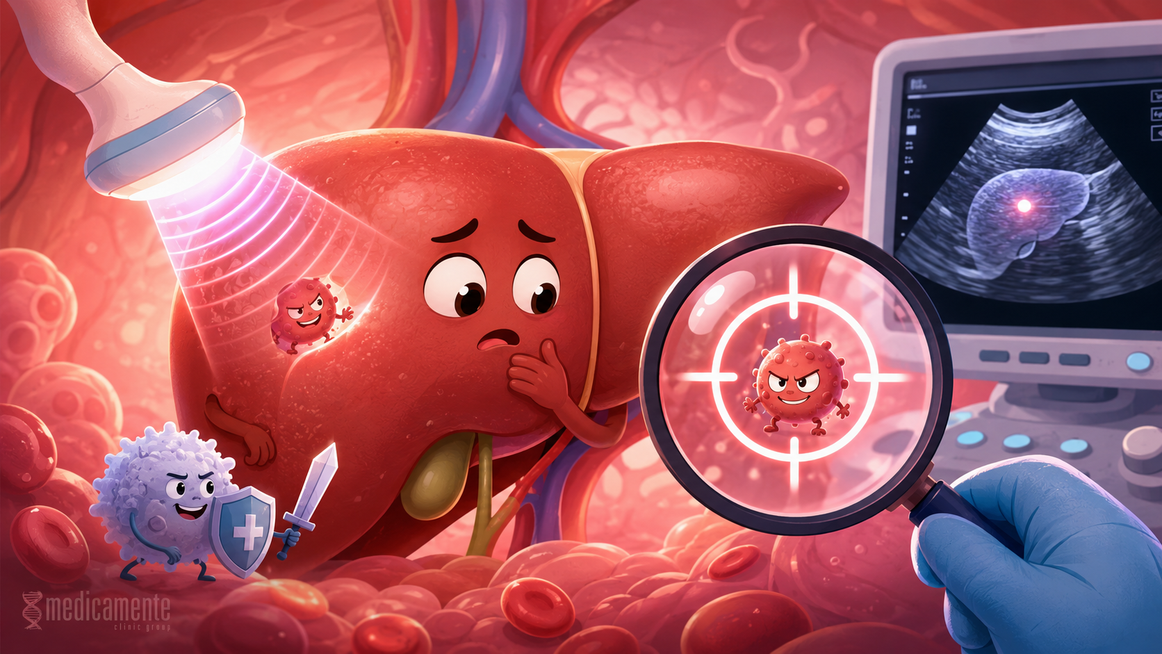
Why chronic liver diseases require regular follow-up
Liver cancer: why it is often connected with chronic diseases
Liver cancer is not a single disease, but a group of malignant tumors that may develop in liver tissue or reach the liver as metastases from other organs. When primary liver cancer is discussed, the term most often refers to hepatocellular carcinoma. It develops from hepatocytes, the main cells of the liver. This type of tumor is closely connected with chronic liver diseases, long-term inflammation, fibrosis and cirrhosis.
The liver has a high capacity for recovery. If damage is short-term, liver cells can regenerate and the structure of the organ is preserved. But in chronic inflammation, this process becomes continuous. Cells are damaged, die, recover and are again exposed to viruses, fat, toxins, immune inflammation or metabolic dysfunction. The longer this cycle of injury and regeneration continues, the higher the likelihood of errors during cell division and accumulation of changes that may lead to tumor growth.
This is why hepatocellular carcinoma often develops not in a completely healthy liver, but against the background of chronic disease. The most important risk factors include cirrhosis, chronic hepatitis B, chronic hepatitis C, hepatitis D, metabolic dysfunction-associated fatty liver disease, alcohol-related injury, inherited metabolic diseases and some autoimmune liver diseases. In many patients, liver cancer is not the first manifestation of disease, but a late complication of a long-lasting process.
Why cirrhosis increases tumor risk
Cirrhosis is one of the main risk factors for primary liver cancer. In cirrhosis, normal organ structure is replaced by scar tissue and regenerative nodules. Inside these nodules, liver cells continue to divide, trying to maintain organ function. But the surrounding tissue is already altered: blood flow is disrupted, chronic inflammation is present, the immune environment changes and conditions are created for the accumulation of genetic abnormalities.
The longer cirrhosis exists, the greater the need for regular follow-up. It is important to understand that the risk of liver cancer remains even when the cause of cirrhosis has already been brought under control. For example, after successful treatment of hepatitis C or effective suppression of hepatitis B virus, the risk may decrease, but if cirrhosis has already formed, it does not disappear completely. Therefore, patients with cirrhosis need systematic monitoring regardless of how they feel.
A key feature of hepatocellular carcinoma is that it may develop for a long time without obvious symptoms. While the tumor is small, a person may not feel pain, pronounced weakness, jaundice or weight loss. If symptoms appear, this often means that the tumor has already reached a significant size, impaired liver function or spread beyond the initial stage. This is why early detection of liver cancer cannot be based only on waiting for complaints.
Viral hepatitis and the risk of liver cancer
Chronic viral hepatitis plays an important role in the development of liver cancer. Hepatitis B is especially significant because it may increase the risk of hepatocellular carcinoma even before advanced cirrhosis has formed. This is related to the specific interaction of the virus with liver cells, long-term inflammation and the influence of viral components on cellular processes. Therefore, patients with chronic HBV require individual risk assessment, even if liver function remains preserved for a long time.
Hepatitis C more often leads to liver cancer through long-term inflammation, fibrosis and cirrhosis. After successful antiviral treatment, the risk decreases, but in patients with already formed advanced fibrosis or cirrhosis, the need for surveillance remains. This is one of the important principles of modern hepatology: eliminating the cause of damage does not always mean complete disappearance of all long-term risks if liver structure has already been significantly altered.
Hepatitis D is considered a particularly aggressive infection because it exists only in the presence of hepatitis B, but it can significantly accelerate liver damage. In patients with HBV/HDV coinfection, fibrosis and cirrhosis develop faster, and the risk of tumor complications increases along with them. Therefore, detecting HDV in patients with hepatitis B is important not only for treatment selection, but also for assessing long-term oncological risk.
Fatty liver disease and a new structure of risk
In recent years, liver cancer associated with metabolic disorders has become increasingly important. Metabolic dysfunction-associated fatty liver disease has become one of the most common causes of chronic liver damage. It is linked to obesity, type 2 diabetes, insulin resistance, lipid metabolism disorders and chronic low-grade inflammation.
If fatty liver disease progresses to steatohepatitis, inflammation and hepatocyte injury occur. In some patients, fibrosis gradually develops, then cirrhosis, and against this background the risk of hepatocellular carcinoma increases. However, an important feature of metabolic liver disease is that a tumor may sometimes develop even without already established cirrhosis. This makes the problem more complex because not all patients with fatty liver disease clearly belong to an obvious high-risk group.
Patients with type 2 diabetes, pronounced obesity, visceral fat, long-term inflammation and signs of fibrosis have a higher risk. Therefore, in metabolic liver diseases, it is important not to stop at the phrase “fat in the liver,” but to assess the degree of fibrosis, associated factors and the need for dynamic follow-up.
Who especially needs follow-up
Early detection of liver cancer is most important in patients from increased-risk groups. This does not mean that every person without risk factors needs frequent liver screening for tumors. But when risk is elevated, surveillance becomes part of medical management.
Increased-risk groups include:
- Patients with cirrhosis of any cause.
Cirrhosis remains one of the main indications for regular surveillance for hepatocellular carcinoma. - Patients with chronic hepatitis B.
For some patients, surveillance may be indicated even without cirrhosis, especially when additional risk factors are present. - Patients after hepatitis C with advanced fibrosis or cirrhosis.
Even after successful treatment of the virus, structural liver changes may preserve oncological risk. - Patients with hepatitis D.
This infection can accelerate liver damage and increase the risk of severe outcomes. - Patients with metabolic liver disease and advanced fibrosis.
This is especially important when combined with type 2 diabetes, obesity, arterial hypertension and dyslipidemia. - Patients with inherited metabolic diseases.
Hemochromatosis, Wilson disease and some other conditions may increase the risk of chronic liver damage. - Patients with a family history of liver cancer.
Cases of hepatocellular carcinoma in close relatives may influence risk assessment.
Follow-up should be prescribed individually. The physician evaluates not only the diagnosis, but also age, cause of disease, degree of fibrosis, test results, family history, comorbidities and the overall condition of the liver.
Which methods are used for early detection
The foundation of surveillance is usually regular visualization of the liver. The most accessible and commonly used method remains ultrasound examination. Ultrasound makes it possible to assess liver structure, detect focal lesions, signs of cirrhosis, ascites, spleen enlargement and other changes. Its advantages include safety, availability and the possibility of repeated use.
However, ultrasound has limitations. The quality of the examination depends on the experience of the specialist, body habitus, degree of obesity, intestinal gas, liver structure and tumor size. In patients with pronounced steatosis or severe cirrhotic remodeling, detection of small lesions may be more difficult. Therefore, if findings are suspicious or ultrasound is insufficiently informative, additional methods may be required.
CT and MRI are used to clarify the nature of a focal lesion. These methods allow assessment of the blood supply characteristics of a nodule, its structure, size, extent and relationship to vessels. For hepatocellular carcinoma, the pattern of contrast enhancement in different phases of imaging is especially important. In some cases, the diagnosis may be established based on a typical CT or MRI pattern without biopsy, if the patient belongs to a high-risk group.
Alpha-fetoprotein is a laboratory marker often used together with imaging methods. Its elevation may accompany hepatocellular carcinoma, but this marker is not ideal. In some patients with liver cancer it remains normal, while in others it may increase during active inflammation or other conditions. Therefore, alpha-fetoprotein should not be considered an independent diagnostic method. It is useful only in combination with the clinical picture and imaging.
Why the early stage is decisive
It is critically important to detect liver cancer early because treatment options strongly depend on stage. If the tumor is small, limited to the liver and organ function is preserved, methods with potentially radical intent may be considered. These include surgical removal of the tumor, liver transplantation in selected patients, local ablation and other local methods.
If the disease is detected late, when the tumor is large, multiple, invades blood vessels or spreads beyond the liver, treatment options become more limited. In such cases, systemic drugs, locoregional interventions, methods of tumor growth control and supportive therapy are used. They may improve prognosis and control the disease, but early detection usually provides more options.
An important feature of liver cancer is that treatment depends not only on tumor size, but also on the condition of the liver itself. A patient may have a small tumor but severe decompensated cirrhosis, which limits surgical options. Or, conversely, liver function may be preserved, allowing a more active strategy to be considered. Therefore, in hepatocellular carcinoma, two processes are always assessed simultaneously: the tumor and the condition of the liver.
Modern approaches to treatment
Treatment of liver cancer has become more diverse. It includes surgical, local, transplantation-based and systemic methods. The choice depends on tumor stage, number of lesions, vessel involvement, liver function, presence of cirrhosis, the patient’s general condition and comorbidities.
The main treatment directions include:
- Surgical removal of the tumor.
This may be used when the lesion is limited and liver function allows safe removal of part of the organ. - Liver transplantation.
In selected patients with cirrhosis and limited tumor burden, transplantation addresses two problems at once: it removes the tumor and replaces the diseased liver. - Local ablation.
Methods that destroy the tumor using heat, cold or other effects may be used for small lesions. - Locoregional therapy.
This includes methods that act on the tumor through liver vessels, such as embolization procedures. - Systemic therapy.
Modern drugs can affect tumor growth, vascular mechanisms and immune response. They are used in advanced disease or when local methods are not possible. - Supportive treatment and control of complications.
In patients with cirrhosis, it is important to treat the tumor and support liver function at the same time.
Modern therapy for hepatocellular carcinoma is developing, but it does not cancel the main principle: the earlier the tumor is detected, the more possibilities there are for effective treatment.
What can be done to reduce risk
Prevention of liver cancer begins with prevention and treatment of chronic liver diseases. If the cause of damage is removed or controlled, the likelihood of progression to fibrosis, cirrhosis and tumor complications can be reduced.
Important measures include:
- Vaccination against hepatitis B.
- Timely detection and treatment of viral hepatitis.
- Control of fatty liver disease and metabolic risk factors.
- Avoidance of alcohol in chronic liver diseases.
- Control of diabetes, body weight, lipid levels and blood pressure.
- Diagnosis and treatment of autoimmune and cholestatic liver diseases.
- Regular surveillance in cirrhosis and advanced fibrosis.
- Cautious use of medications and supplements that may damage the liver.
It is important to understand that prevention of liver cancer is not one blood test and not one examination. It is a long-term strategy for preserving liver health. It begins long before a tumor appears: at the stage of vaccination, hepatitis diagnosis, weight control, diabetes treatment, fibrosis assessment and regular follow-up of patients from risk groups.
Main conclusion
Liver cancer most often develops not suddenly, but against the background of long-term chronic damage to the organ. Cirrhosis, viral hepatitis, fatty liver disease, autoimmune and inherited disorders can create conditions in which the risk of hepatocellular carcinoma increases. At the same time, early tumor stages often do not cause symptoms, so waiting for complaints is an unreliable strategy.
Modern early detection is based on risk group assessment, regular liver imaging, laboratory markers and timely clarification of suspicious lesions using CT or MRI. The main goal of surveillance is to detect a tumor at a stage when the most effective treatment methods are still possible.
The liver can compensate for damage for a long time, but this feature is exactly what makes chronic liver diseases deceptive. A patient may feel well while structural changes are already occurring in the organ. Therefore, regular follow-up in cirrhosis, viral hepatitis and advanced fibrosis is not a formality, but one of the key ways to reduce the risk of late detection of liver cancer.
Write a review
Required fields are marked with *
Categories
- Therapy (81)
- News (48)
- GP (23)
- Cardiology (20)
- urology (12)
- Endocrinology (8)
- Ortopedics (4)
- Dermatology (3)
- Ultrasound (1)
- Mobile app (1)
Articles





Archive
- September 2026 (11)
- August 2026 (11)
- July 2026 (11)
- June 2026 (11)
- May 2026 (9)
- April 2026 (8)
- March 2026 (8)
- February 2026 (8)
- January 2026 (8)
- December 2025 (5)
Categories
- Therapy (81)
- News (48)
- GP (23)
- Cardiology (20)
- urology (12)
- Endocrinology (8)
- Ortopedics (4)
- Dermatology (3)
- Ultrasound (1)
- Mobile app (1)


