
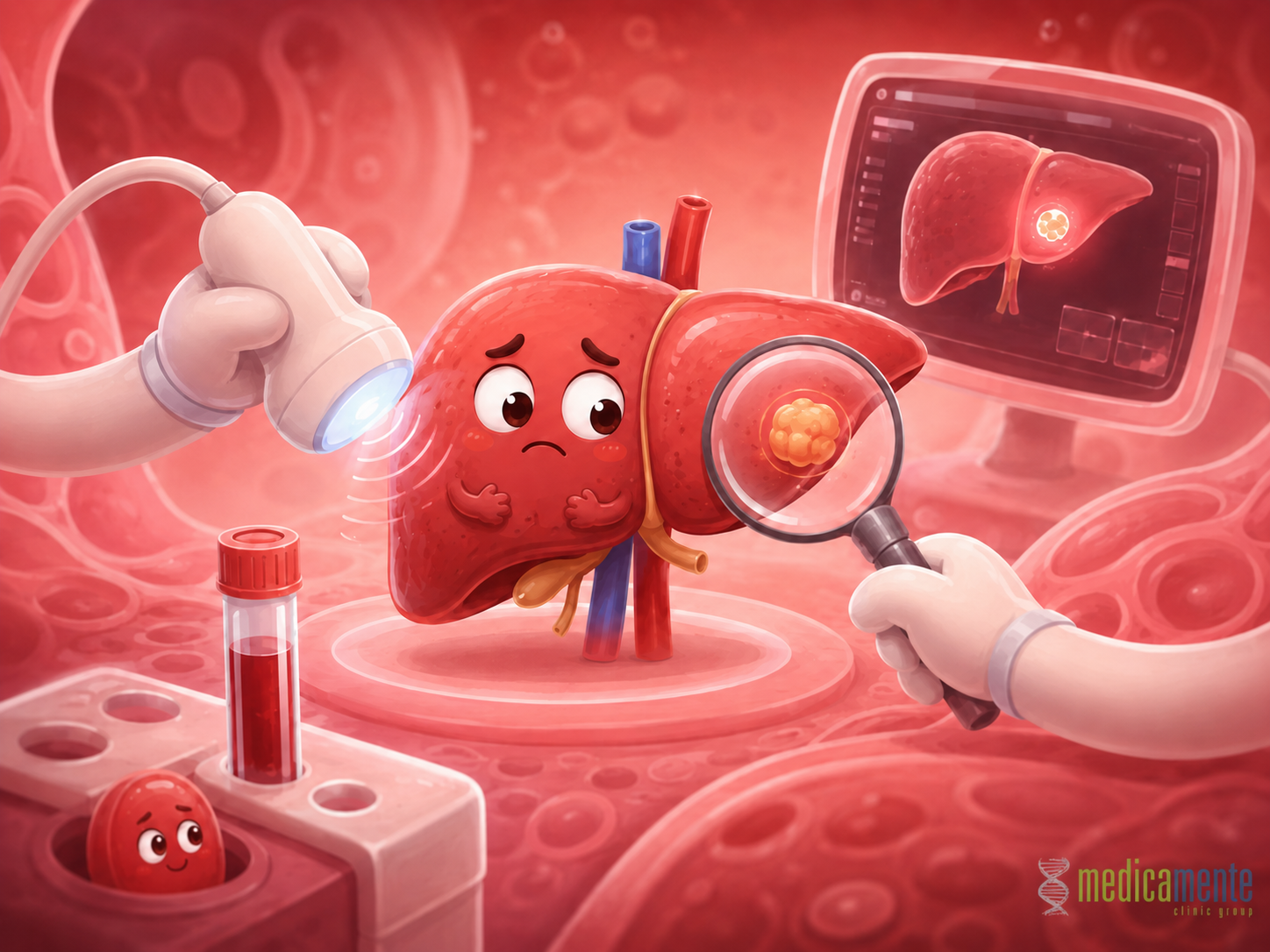
From liver tests to elastography and MRI
Why liver diagnosis requires a comprehensive approach
Liver diseases often develop silently. This is related to the organ’s high compensatory capacity: even with inflammation, fat accumulation, fibrosis or early structural changes, a person may not feel pronounced symptoms for a long time. Pain in the liver area is not a reliable sign because liver tissue itself does not have the same pain sensitivity as skin or muscles. Therefore, the absence of pain does not mean that the liver is completely healthy.
Modern liver diagnosis is based not on one marker, but on a combination of findings. The physician evaluates symptoms, medical history, risk factors, laboratory results, virological tests, immunological markers, instrumental examinations and the dynamics of changes over time. This approach is important because different liver diseases may produce similar abnormalities in blood tests, but require completely different management.
For example, elevation of ALT and AST may occur in viral hepatitis, fatty liver disease, autoimmune hepatitis, drug-induced liver injury, toxic exposure and some metabolic diseases. Elevation of GGT and alkaline phosphatase may indicate impaired bile flow, but it also requires clarification of the cause. Therefore, the task of diagnosis is not simply to find an abnormal result, but to understand its origin, severity and possible consequences for the patient.
Liver tests: what the main markers show
Liver tests are a group of laboratory markers that help assess liver cell injury, impaired bile flow and some functions of the organ. They often become the first stage of diagnosis, but they do not provide a complete answer on their own.
The main markers include:
- ALT and AST.
These enzymes increase when liver cells are damaged. ALT is considered more specific to the liver, while AST may also increase with injury to muscles, the heart and other tissues. It is important to assess not only the fact of elevation, but also its degree, dynamics and combination with other markers. - GGT and alkaline phosphatase.
These markers are often associated with cholestasis, meaning impaired bile formation or bile flow. They may increase in diseases of the bile ducts, drug-related effects, alcohol-related injury, fatty liver disease and other conditions. - Bilirubin.
Bilirubin reflects bile pigment metabolism. Its elevation may appear as yellowing of the skin and sclera, dark urine and changes in stool color. The causes may differ: liver injury, impaired bile flow, hemolysis or inherited features of metabolism. - Albumin.
Albumin reflects the synthetic function of the liver. In advanced chronic liver disease, its level may decrease, showing reduced ability of the organ to produce important proteins. - INR and prothrombin time.
These markers help assess the blood clotting system. The liver synthesizes many clotting factors, so severe impairment of its function may lead to coagulation abnormalities. - Platelets.
A decrease in platelet count may be an indirect sign of portal hypertension and spleen enlargement in advanced fibrosis or cirrhosis.
It is important to understand that normal liver tests do not always completely exclude liver disease. In some patients with fatty liver disease, chronic viral hepatitis or fibrosis, markers may be normal or change only intermittently. Therefore, laboratory results must be interpreted in the context of risk factors and imaging findings.
Medical history and risk factors: why the physician’s questions matter
Liver diagnosis begins not only with the laboratory. A detailed medical history often helps determine the direction of further evaluation. The physician asks about previous viral infections, vaccination, surgeries, blood transfusions, injection procedures, medication use, alcohol intake, dietary habits, body weight, diabetes, family diseases, itching, jaundice, stool and urine changes and possible autoimmune diseases.
Risk factors may point to the likely cause of liver damage. For example, type 2 diabetes, obesity, high triglyceride levels and arterial hypertension make metabolic dysfunction-associated fatty liver disease more likely. Contact with blood, lack of vaccination and certain epidemiological factors require exclusion of viral hepatitis. Autoimmune diseases in the patient or family may increase the likelihood of autoimmune liver injury.
Medications and dietary supplements must also be considered. Some drugs can damage the liver, especially with long-term use, combination of several medications or the presence of an already existing liver disease. Herbal products and supplements are not always safe either. Therefore, when liver tests are abnormal, it is important to list honestly everything the patient takes regularly or has taken in recent months.
Virological diagnosis
Viral hepatitis remains one of the key causes of chronic liver disease. Therefore, when liver injury is suspected, testing for hepatitis B, C and D is often required. These infections may remain asymptomatic for a long time, but gradually lead to fibrosis, cirrhosis and an increased risk of liver cancer.
In hepatitis B, diagnosis includes detection of HBsAg, antibodies to different viral antigens, HBV DNA and other markers when indicated. These tests help determine whether there is a current infection, whether the person had hepatitis in the past, whether immunity has formed after vaccination or contact with the virus, how active viral replication is and whether treatment is needed.
In hepatitis C, antibodies to HCV and detection of HCV RNA are important. Antibodies show whether there has been contact with the virus, but they do not always mean active infection. To confirm an ongoing viral process, viral RNA must be detected. This matters because modern hepatitis C therapy allows most patients to achieve a sustained virological response.
Hepatitis D is tested in patients with confirmed hepatitis B because virus D cannot exist without HBsAg. Diagnosis includes antibodies to HDV and detection of HDV RNA when necessary. This is especially important if a patient with HBV has rapid fibrosis progression, persistent liver inflammation or signs of a more aggressive disease course.
Immunological and metabolic tests
Not all liver diseases are connected with viruses or metabolic disorders. In some patients, the cause is an autoimmune process. In such cases, the immune system damages liver cells or bile ducts. Autoantibodies, immunoglobulins and additional investigations may be used for diagnosis.
When autoimmune hepatitis is suspected, the physician may evaluate autoantibodies, IgG level, liver enzyme activity and exclude other causes of inflammation. In primary biliary cholangitis, markers of cholestasis, antimitochondrial antibodies and IgM level are important. When primary sclerosing cholangitis is suspected, signs of bile duct involvement and imaging methods become more significant.
Inherited and metabolic liver diseases have a separate place. When hemochromatosis is suspected, iron metabolism markers are evaluated. When Wilson disease is suspected, copper metabolism is examined. In some conditions, genetic tests, special biochemical investigations and consultation with a specialist may be required. These diseases are less common, but it is important not to miss them, especially in young patients or in an unusual pattern of liver injury.
Liver ultrasound: an accessible method of primary imaging
Ultrasound examination often becomes the first instrumental method for assessing the liver. It helps visualize organ size, tissue structure, signs of fatty infiltration, focal lesions, bile duct dilatation, gallbladder condition, fluid in the abdominal cavity and indirect signs of portal hypertension.
Ultrasound is convenient because it is safe, accessible and can be repeated over time. In fatty liver disease, it may reveal signs of steatosis. In cirrhosis, it may show changes in liver contours, heterogeneous structure, spleen enlargement, ascites and other signs. When focal lesions are suspected, ultrasound may be the first method that detects a nodule.
However, ultrasound has limitations. It depends on the quality of equipment, the specialist’s experience, the patient’s body habitus, intestinal gas, degree of obesity and liver structure. Small focal lesions may be difficult to detect, especially in pronounced steatosis or cirrhosis. Therefore, if suspicious findings are present, CT or MRI may be required.
Elastography: fibrosis assessment without biopsy
Elastography has become an important method in modern hepatology. It allows assessment of liver tissue stiffness, which increases in fibrosis. The more connective tissue accumulates in the liver, the higher the stiffness. This helps estimate the likelihood of significant fibrosis or cirrhosis without an invasive procedure.
The method is especially useful in chronic viral hepatitis, fatty liver disease, alcohol-related injury and other chronic conditions. Elastography helps determine how far the disease has progressed, how often follow-up is needed and whether more active treatment is required. It can also be used over time to monitor changes during therapy or after removal of the damaging factor.
But elastography is not an absolute method either. The result may be influenced by active inflammation, congestion in the liver, cholestasis, food intake before the examination, pronounced obesity and technical factors. Therefore, the stiffness value must be interpreted together with blood tests, medical history, ultrasound and the clinical picture.
CT, MRI and specialized imaging methods
Computed tomography and magnetic resonance imaging are used when liver structure, vessels, focal lesions or bile ducts need more precise assessment. These methods are especially important when liver tumor, complex cysts, vascular lesions, complications of cirrhosis or unclear ultrasound findings are suspected.
Contrast-enhanced liver MRI can provide detailed information about the nature of a lesion, blood supply characteristics and tissue structure. CT is also widely used to assess tumors, vascular anatomy, disease extent and complications. The choice between CT and MRI depends on the clinical task, contraindications, availability of the method and the patient’s condition.
MR cholangiography may be used to assess the bile ducts. It helps detect strictures, dilatations, stones, inflammatory changes or signs of sclerosing cholangitis. In selected cases, endoscopic methods are used; they may be not only diagnostic, but also therapeutic, for example when stones are removed or bile duct obstruction is relieved.
Liver biopsy: when it is still needed
Despite the development of non-invasive methods, liver biopsy remains important in selected clinical situations. It makes it possible to obtain a tissue fragment and examine it under a microscope. This may be important when the diagnosis remains unclear, laboratory tests and imaging give contradictory information, or it is necessary to determine precisely the activity of inflammation, degree of fibrosis, signs of steatohepatitis, autoimmune process or drug-induced injury.
Biopsy is not performed in every patient. It is an invasive procedure, so the decision must be justified. The physician evaluates whether the biopsy result will change treatment strategy. If the answer can be obtained through blood tests, elastography and imaging, biopsy may not be needed. But if the diagnosis remains uncertain without morphological confirmation, this method remains important.
How examination results are combined
The main mistake in liver diagnosis is to evaluate each marker separately. Elevated ALT is not a diagnosis. Fatty infiltration on ultrasound does not automatically indicate the degree of fibrosis. Normal bilirubin does not exclude chronic liver disease. Increased stiffness on elastography requires comparison with inflammation, cholestasis and other factors.
Correct diagnosis answers several questions:
- Is there liver damage.
- Which mechanism predominates: liver cell inflammation, cholestasis, fat accumulation, fibrosis or a mixed pattern.
- Which cause is most likely.
- Are there signs of advanced fibrosis or cirrhosis.
- Is liver function preserved.
- Is there a risk of complications.
- Is treatment, follow-up or additional examination needed.
This approach allows the physician not simply to describe laboratory abnormalities, but to build a clinical picture. This is what distinguishes complete diagnosis from a random set of investigations.
Main conclusion
Modern diagnosis of liver diseases has become much more precise than before. It includes liver tests, virological studies, immunological markers, assessment of metabolic factors, ultrasound, elastography, CT, MRI and, when necessary, biopsy. But the main principle remains unchanged: no method should be evaluated separately from the patient.
The liver can compensate for damage for a long time, so diseases are often detected not by symptoms, but through laboratory and instrumental findings. This makes preventive examination and risk factor assessment especially important. The earlier the cause of damage is identified, the greater the possibility of slowing progression, preventing fibrosis, reducing the risk of cirrhosis and detecting complications in time.
Liver diagnosis is not the search for one “main test,” but a step-by-step understanding of the organ’s condition. The physician must determine the cause, activity of the process, degree of structural change and prognosis. This approach makes it possible to move from accidental detection of abnormalities to conscious patient management and preservation of liver function over the long term.
Write a review
Required fields are marked with *
Categories
- Therapy (81)
- News (48)
- GP (23)
- Cardiology (20)
- urology (12)
- Endocrinology (8)
- Ortopedics (4)
- Dermatology (3)
- Ultrasound (1)
- Mobile app (1)
Articles





Archive
- September 2026 (11)
- August 2026 (11)
- July 2026 (11)
- June 2026 (11)
- May 2026 (9)
- April 2026 (8)
- March 2026 (8)
- February 2026 (8)
- January 2026 (8)
- December 2025 (5)
Categories
- Therapy (81)
- News (48)
- GP (23)
- Cardiology (20)
- urology (12)
- Endocrinology (8)
- Ortopedics (4)
- Dermatology (3)
- Ultrasound (1)
- Mobile app (1)


