How water, salts, and metabolism affect recurrence risk
Kidney stones: why this is not always a one-time problem
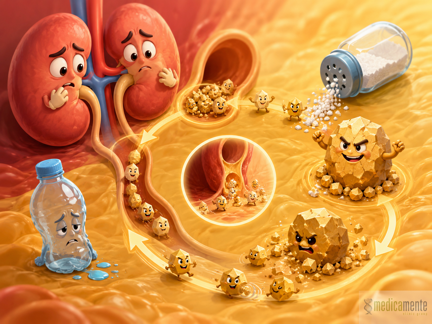
Kidney stones form when substances in the urine accumulate and begin to create crystals. If these conditions persist long enough, crystals can grow, combine with each other, and form a stone. This process may occur in the kidneys, ureters, or other parts of the urinary system.
Many patients perceive a kidney stone as a one-time event: pain appears, the stone passes or is removed, and the problem seems finished. However, kidney stone disease often tends to recur. If the reason for stone formation is not addressed, new stones may appear again months or years later.
The risk of recurrence depends on the composition of the stone, fluid intake, urine concentration, diet, infections, urine acidity, calcium metabolism, oxalates, uric acid, citrates, and other factors. After an episode of kidney stone disease, it is therefore important not only to remove the specific stone, but also to understand why it formed.
How stones form
Urine contains many dissolved substances: calcium salts, oxalates, phosphates, uric acid, sodium, potassium, magnesium, citrates, and other compounds. Normally, they remain dissolved and are excreted from the body. But if their concentration becomes too high or urine acidity changes, crystals may begin to form.
Stone formation depends on the balance between substances that promote crystallization and substances that inhibit it. For example, citrates help prevent the formation of some stones. If citrate levels are low, while calcium or oxalate levels are high, the risk of crystallization increases.
Urine volume plays an important role. If a person drinks too little fluid, loses a lot of water through sweating, or is often exposed to heat, urine becomes more concentrated. In concentrated urine, salts more easily reach levels at which crystals begin to form. This is why insufficient fluid intake is one of the clearest and most common risk factors.
However, stones do not form only because of water intake. Some people have metabolic features such as increased urinary excretion of calcium, oxalates, or uric acid, low citrate levels, changes in urine pH, hereditary predisposition, or diseases that affect metabolism. In such cases, the general advice to “drink more water” may not be enough.
Types of kidney stones
Kidney stones differ in composition. This is important because prevention depends on the type of stone. The same approach does not work for all patients.
Calcium stones are the most common. They are most often made of calcium oxalate, less often calcium phosphate. These stones may be associated with increased urinary calcium or oxalate excretion, low citrate levels, low urine volume, dietary factors, and certain metabolic disorders.
Uric acid stones are made of uric acid. They form more often in acidic urine. The risk is higher with elevated uric acid, gout, metabolic syndrome, obesity, diabetes, high purine intake, and low urine volume. A special feature of uric acid stones is that in some cases they may be dissolved by proper correction of urine pH, but this should be done under medical supervision.
Struvite stones are associated with urinary tract infections, especially bacteria that change urine composition and make it more alkaline. These stones can grow quickly and become large. In such cases, treating the infection and evaluating anatomical or functional reasons for recurrence are especially important.
Cystine stones are less common and are related to a hereditary disorder of amino acid metabolism. They may appear at a young age and tend to recur. These cases require specialized follow-up.
If a stone passes naturally or is removed, its analysis can provide valuable information. Determining stone composition helps choose prevention more accurately than general recommendations.
Why stones cause severe pain
A stone may remain in the kidney for a long time without causing symptoms. But if it starts moving and enters the ureter, it can block urine flow. The ureter is a narrow tube through which urine travels from the kidney to the bladder. If a stone becomes stuck, pressure above the obstruction increases, the ureter spasms, and renal colic develops.
Renal colic usually presents as sharp pain in the side or lower back, which may radiate to the groin, lower abdomen, or external genital area. The pain is often wave-like and intense, and may be accompanied by nausea, vomiting, frequent urges to urinate, blood in the urine, and marked restlessness.
Pain intensity does not always reflect stone size. A small stone can cause severe colic if it blocks the ureter. A larger stone may remain in the kidney for a long time and cause minimal symptoms if it does not obstruct urine flow.
Particular attention is needed when pain is accompanied by high fever, chills, marked weakness, reduced urine output, a single functioning kidney, pregnancy, or known chronic kidney disease. In such situations, the risk of complications is higher.
Why stones may form again
Recurrent stone formation occurs when the conditions for crystallization remain. The stone itself may be removed, but metabolic or behavioral factors remain unchanged. Recurrence is therefore often related not to unsuccessful treatment of a specific episode, but to the lack of prevention afterward.
One common reason is insufficient urine volume. If urine is constantly concentrated, salts more easily precipitate into crystals. This is especially relevant for people who drink little fluid, sweat a lot, do physical work, live in a hot climate, or often restrict fluids because they are busy.
Diet can also influence risk. Excessive salt intake increases urinary calcium excretion and may contribute to calcium stone formation. A large amount of foods high in oxalates may matter in people prone to oxalate stones. High purine intake may increase the risk of uric acid stones. However, preventive diet should be individualized because different stones require different approaches.
Metabolic factors include increased urinary calcium, oxalate, or uric acid excretion, low citrate levels, changes in urine acidity, obesity, insulin resistance, gout, bowel diseases, malabsorption, and some hereditary conditions.
Urinary tract infections may also support stone formation. This is especially true for struvite stones, which are associated with bacteria that change the chemical environment of urine. If infection persists or recurs frequently, the risk of such stones increases.
Which tests help identify the cause
After the first episode of kidney stone disease, the extent of evaluation depends on the situation. Some patients need only urinalysis, blood tests, imaging, and basic risk assessment. Others need expanded metabolic testing, especially if stones recur, appear at a young age, have an unusual composition, or are associated with chronic kidney disease, infections, or family history.
Common tests may include:
- Urinalysis.
It helps detect blood, white blood cells, bacteria, crystals, urine pH, and specific gravity. - Blood tests.
Creatinine, calcium, uric acid, electrolytes, and other markers may be assessed when indicated. - Kidney and urinary tract ultrasound.
It can detect stones, urinary tract dilation, signs of impaired urine flow, cysts, and other changes. - CT when a stone is suspected.
Computed tomography is often used to accurately detect stones and assess their size, location, and density. - Stone composition analysis.
If the stone is available, laboratory analysis helps determine its type and choose prevention. - 24-hour urine testing.
In recurrent stones or high-risk patients, urine volume, calcium, oxalate, uric acid, citrate, sodium, and other parameters may be assessed. - Urine culture.
It is ordered when an infectious stone origin or recurrent infections are suspected.
The goal of evaluation is not only to confirm the presence of a stone, but also to understand what conditions led to its formation and what can be changed.
The role of water and fluid intake
Adequate fluid intake is one of the basic elements of kidney stone prevention. The higher the urine volume, the lower the concentration of salts and the lower the chance of crystallization. This does not mean that the same amount of water is suitable for every patient. Fluid needs depend on body weight, physical activity, climate, sweating, diet, heart function, and kidney function.
A practical sign is that urine should not be constantly dark and concentrated. If a person urinates rarely, urine is intensely colored, and tests show high specific gravity, this may suggest insufficient fluid intake. However, in heart failure, advanced chronic kidney disease, or a tendency to swelling, fluid intake should be discussed with a doctor.
It is especially important to increase fluid intake when water loss increases: heat, physical exertion, fever, work in dry environments, or active training. In these situations, the risk of concentrated urine is higher.
Not all drinks are equally useful. Sugary drinks, excessive carbonated beverages, and large amounts of alcohol are not good preventive strategies. In most cases, ordinary fluid intake should be the foundation, while specific recommendations depend on stone type and the patient’s condition.
Diet in people prone to stones
Diet in kidney stone disease should take stone composition into account. A universal strict diet without understanding the cause may be useless or even incorrect. For example, in calcium stones, calcium intake from food should not always be sharply restricted. Too little dietary calcium may increase oxalate absorption in the intestine and increase oxalate excretion in urine.
Salt is one of the most important factors. Excessive sodium intake can increase urinary calcium excretion. Reducing salt intake is therefore often recommended in patients prone to calcium stones and hypertension. It is important to consider hidden salt in ready-made foods, sauces, processed meats, cheeses, semi-prepared foods, and fast food.
In oxalate stones, moderate restriction of foods high in oxalates may be relevant if tests confirm increased oxalate excretion. Such foods may include spinach, sorrel, rhubarb, certain nuts, cocoa, and other products. However, large groups of foods should not be excluded without indication.
In uric acid stones, more attention is paid to uric acid, urine pH, body weight, metabolic syndrome, and purine intake. In such cases, excessive intake of red meat, organ meats, and some other purine-rich foods may be limited. But here too, the approach should be individualized.
Extreme measures should also be avoided: fasting, crash diets, excessive use of protein supplements, and self-administration of high-dose vitamin C or mineral preparations without indication. These actions may affect urine composition and increase stone risk in predisposed people.
Infections and stones
Urinary tract infections can be not only a consequence of stones, but also a cause of their formation. Some bacteria change the chemical composition of urine, making it more alkaline and creating conditions for struvite stones. These stones may grow quickly and sometimes form large kidney stones.
If a stone is infection-related, it is important not to focus only on antibiotics or stone removal. The reason why infection recurs must also be understood. Causes may include residual stone fragments, impaired urine flow, anatomical features, incomplete bladder emptying, diabetes, or other factors.
Infection in the setting of urinary obstruction can be dangerous. If a stone blocks urine flow and infection develops at the same time, the risk of severe complications increases. Therefore, the combination of side pain, fever, chills, and worsening general condition requires urgent medical assessment.
Treatment of stones: from observation to procedures
Treatment strategy depends on stone size, location, composition, symptoms, presence of infection, obstruction of urine flow, kidney condition, and the patient’s overall health. Small stones may sometimes pass on their own, especially if they are located in the ureter and do not cause complications. In such cases, the doctor may choose observation, pain control, monitoring of stone passage, and repeat imaging.
If the stone is large, causes severe pain, does not pass on its own, blocks urine flow, is associated with infection, or threatens kidney function, active treatment may be needed. Different approaches are used: shock wave lithotripsy, endoscopic removal through the urinary tract, percutaneous techniques for large stones, and other procedures.
The choice of method depends on the specific situation. Stone size, density, and location, urinary tract anatomy, the presence of inflammation, and technical possibilities all matter. Treatment of kidney stone disease should therefore be individualized.
After stone removal, prevention remains necessary. Even a perfectly performed procedure does not eliminate metabolic causes if they continue to act. The next step is therefore recurrence risk assessment and selection of preventive measures.
When urgent medical help is needed
Kidney stone disease can present in different ways. Sometimes a stone causes moderate discomfort; sometimes it causes severe renal colic. But some situations require especially prompt medical attention.
These warning signs include high fever, chills, marked weakness, side pain with worsening general condition, absence of urine or sharply reduced urine output, vomiting, inability to drink fluids, large amounts of blood in the urine, pregnancy, a single kidney, known chronic kidney disease, or severe pain that does not improve.
The danger is not only pain. If a stone blocks urine flow, pressure in the urinary system rises. If infection is added, the condition can become severe quickly. In such cases, it is not safe simply to wait for the stone to pass.
Kidney stone disease has two important stages: treatment of the current stone and prevention of a new one. If treatment is limited to pain relief or stone removal without identifying the cause, the risk of recurrence remains. After a stone episode, stone composition analysis, urine and blood evaluation, fluid intake, diet, infections, and metabolic factors are all important.
Write a review
Required fields are marked with *
Categories
- Therapy (49)
- News (48)
- GP (23)
- urology (12)
- Cardiology (9)
- Endocrinology (8)
- Ortopedics (4)
- Dermatology (3)
- Check-up (1)
- Ultrasound (1)
Articles

Archive
- June 2026 (11)
- May 2026 (9)
- April 2026 (8)
- March 2026 (8)
- February 2026 (8)
- January 2026 (8)
- December 2025 (5)
- November 2025 (6)
- October 2025 (6)
- September 2025 (6)
Categories
- Therapy (49)
- News (48)
- GP (23)
- urology (12)
- Cardiology (9)
- Endocrinology (8)
- Ortopedics (4)
- Dermatology (3)
- Check-up (1)
- Ultrasound (1)


Comments (0)