
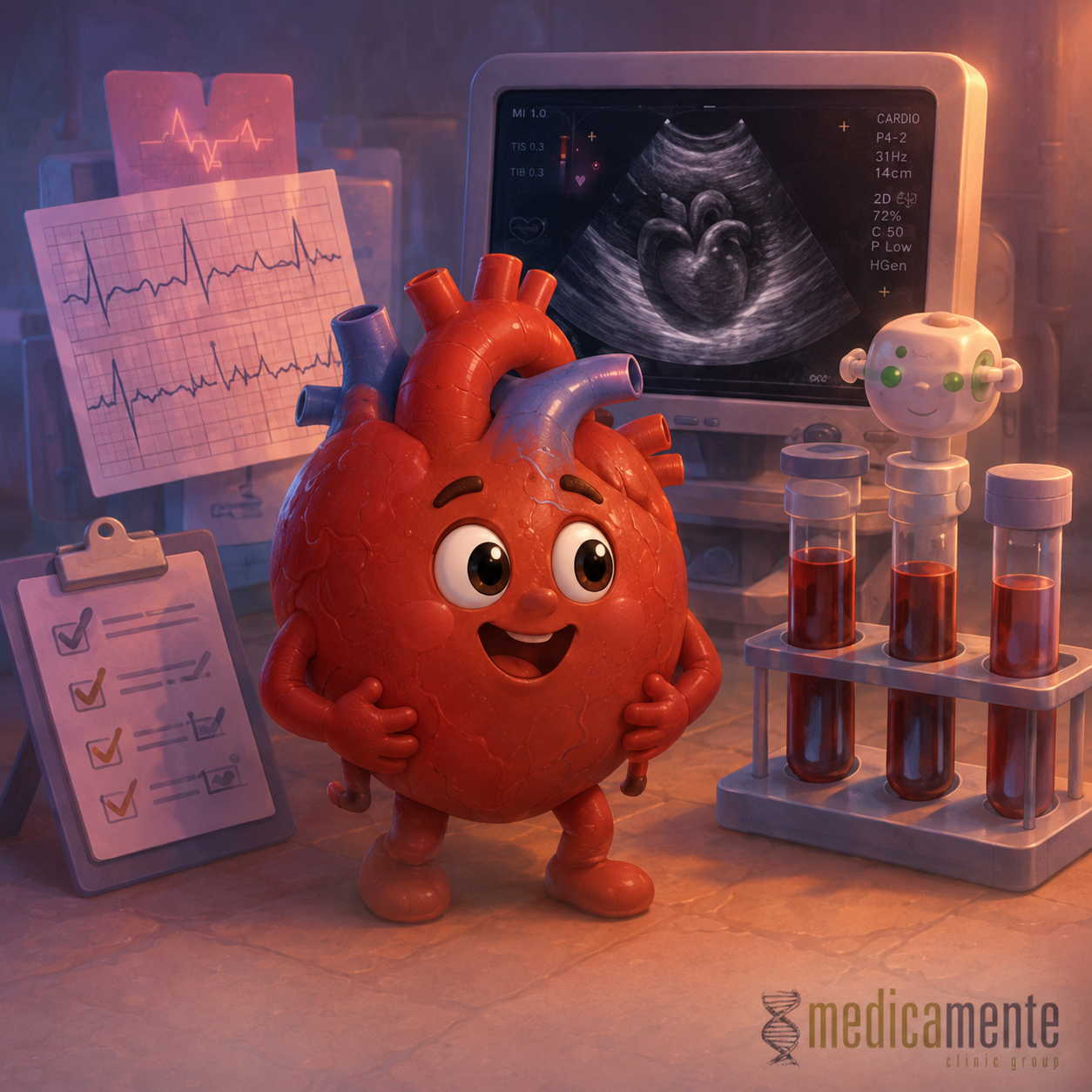
Which tests help assess rhythm, structure and vascular risk
Why the heart cannot be assessed with one test
The heart performs several functions at once. It generates electrical impulses, contracts, pumps blood, works through valves, adapts to pressure and physical activity, receives its own blood supply through the coronary arteries and depends on the condition of the vessels, kidneys, lungs, hormones and metabolism. This is why one test is usually not enough to fully assess the cardiovascular system.
Different methods answer different questions. An electrocardiogram shows the electrical activity of the heart and helps detect rhythm disorders, conduction problems and some signs of ischemia or overload. Echocardiography shows the structure of the heart, the valves, chamber size, contractility and signs of heart failure. Blood tests help assess cholesterol, blood sugar, kidney function, electrolytes, markers of heart injury and overall vascular risk.
Sometimes patients expect a “heart check” to mean one specific test. In practice, the examination is chosen according to the clinical question: whether there is chest pain, shortness of breath, palpitations, fainting, high blood pressure, diabetes, high cholesterol, previous myocardial infarction, suspected arrhythmia or heart failure.
Modern heart diagnostics are based on a step-by-step approach. First, symptoms, risk factors, blood pressure, pulse and basic blood tests are assessed. Then the methods that can confirm or rule out a specific problem are chosen. This helps avoid unnecessary tests while still not missing important disease.
ECG: what an electrocardiogram shows
An electrocardiogram is one of the most accessible and important tests in cardiology. It records the electrical activity of the heart from the surface of the body. With ECG, a doctor assesses rhythm, heart rate, conduction, signs of overload of heart chambers and some changes related to ischemia, previous heart attack or electrolyte disturbances.
ECG is especially important in chest pain, palpitations, fainting, suspected arrhythmia, shortness of breath, high blood pressure and before some types of treatment. In acute situations, ECG can help quickly suspect myocardial infarction or a dangerous rhythm disorder.
However, ECG is a recording at one specific moment. If an arrhythmia occurs in attacks and is not present during the recording, a standard ECG may be normal. This does not mean that the symptoms are imaginary or that there is definitely no problem. In such cases, longer rhythm monitoring may be needed.
It is also important to understand that a normal ECG does not exclude all heart diseases. For example, a person may have atherosclerosis of the coronary arteries or early heart failure while the resting ECG shows no major abnormalities. Therefore, ECG is interpreted together with symptoms, blood tests, blood pressure and other methods.
Holter monitoring
Holter monitoring is a long-term ECG recording, usually over 24 hours or longer. The patient wears a small device that records heart rhythm during normal daily life: walking, sleeping, working, stress and routine activities.
This method is especially useful when symptoms are not constant: skipped beats, palpitations, episodes of weakness, dizziness, near-fainting or irregular pulse. Holter monitoring helps determine whether symptoms match a rhythm disorder, how often extrasystoles occur, whether there are pauses, tachycardia, atrial fibrillation or other arrhythmias.
Holter monitoring also shows how heart rate changes during the day and night. This can be important when very fast or very slow rhythm, conduction disorders, treatment effectiveness or the safety of certain medications needs to be assessed.
If episodes are rare and do not occur during 24-hour monitoring, longer event recorders may be needed. Data from wearable devices can sometimes be useful, but they do not always replace medical diagnostics and require confirmation, especially when important treatment decisions are involved.
Ambulatory blood pressure monitoring
Blood pressure can vary greatly during the day. It depends on physical activity, emotions, sleep, pain, caffeine, medication, stress and even the fact of visiting a doctor. Therefore, a single office measurement does not always reflect the true picture.
Ambulatory blood pressure monitoring assesses blood pressure during the day and night. The patient wears a cuff and a device that automatically takes measurements at set intervals. This helps detect sustained hypertension, nighttime blood pressure elevation, morning surges, excessive blood pressure drops or unstable readings.
This method is especially useful when “white coat hypertension” is suspected, when blood pressure rises during a medical visit but is normal at home. It also helps detect masked hypertension, when office readings are normal but blood pressure is elevated at home or at work.
Nighttime blood pressure is especially important. In many people, blood pressure normally decreases during sleep. If this nighttime decrease is absent or blood pressure rises at night, it may be associated with a higher risk of complications, sleep apnea, kidney disease, diabetes or poor hypertension control.
Echocardiography: ultrasound of the heart
Echocardiography, or heart ultrasound, allows doctors to see the structure and function of the heart in real time. It is one of the main tests used for shortness of breath, suspected heart failure, heart murmurs, valve disease, previous myocardial infarction, arrhythmias, high blood pressure and some congenital or acquired heart diseases.
During echocardiography, chamber size, wall thickness, contractility, ejection fraction, valve function, blood flow direction, pressure in the pulmonary artery and the presence of fluid in the pericardium are assessed. These data help determine whether structural heart disease is present.
Left ventricular ejection fraction is one important measure. It shows what proportion of blood is pumped out of the left ventricle during contraction. A reduced ejection fraction may indicate systolic heart failure. However, a normal ejection fraction does not always exclude heart failure, because the heart may relax poorly or work with elevated filling pressures.
Echocardiography is also important in valve diseases. It helps determine whether a valve is narrowed, whether it closes poorly, whether heart chambers are enlarged and how much strain is placed on the heart muscle. The results may influence the need for follow-up, treatment or cardiac surgery consultation.
Stress tests
Some heart problems appear only during physical activity. At rest, blood supply may be sufficient, but during walking, climbing stairs or exercise, the heart needs more oxygen. If the coronary arteries are narrowed, ischemia may appear.
Stress tests help evaluate how the heart responds to physical activity. Bicycle ergometry or treadmill testing is commonly used, with controlled exercise under ECG, blood pressure and symptom monitoring. The doctor assesses exercise tolerance, chest pain, shortness of breath, ECG changes or rhythm disorders.
Stress tests are not used for everyone. They are useful for specific symptoms and levels of risk, especially when the probability of coronary artery disease or the safety of physical activity needs to be assessed. If the patient has acute symptoms, unstable condition or certain contraindications, a stress test is not performed.
In some cases, stress echocardiography, stress MRI or other imaging methods under stress are used. These methods help show how different parts of the heart contract when oxygen demand increases.
Blood tests: why they matter for the heart
The heart cannot be assessed only with ECG and ultrasound. Blood tests help reveal risk factors and conditions that affect the cardiovascular system. Many of them cause no symptoms but gradually increase the risk of complications.
A lipid profile shows total cholesterol, LDL, HDL and triglycerides. It helps assess the risk of atherosclerosis and the need for correction. LDL cholesterol is especially important because long-term elevation is linked to arterial damage.
Glucose and glycated hemoglobin help detect diabetes, prediabetes and the quality of glucose control. Diabetes significantly increases the risk of heart attack, stroke, heart failure and kidney involvement.
Creatinine and estimated glomerular filtration rate show kidney function. This is important because chronic kidney disease increases cardiovascular risk, affects blood pressure, fluid retention and medication safety. Electrolytes, especially potassium and sodium, are important for heart rhythm and for treating hypertension, heart failure and arrhythmias.
Troponin: a marker of heart muscle damage
Troponin is a protein found in heart muscle cells. When myocardial cells are damaged, troponin can enter the bloodstream. This is why a troponin test is used when myocardial infarction and other conditions involving heart injury are suspected.
It is important to understand that troponin is not a preventive test “just in case.” Its main purpose is to assess a patient with symptoms, such as chest pain, shortness of breath, marked weakness or suspected acute heart disease. The result is interpreted together with ECG, symptoms, timing and changes over time.
Elevated troponin does not always mean a classic heart attack caused by a clot in a coronary artery. It can also increase in other conditions, such as severe heart failure, myocarditis, marked tachycardia, severe infection, kidney failure, pulmonary embolism and other causes. Clinical context is therefore essential.
Sometimes troponin is repeated after several hours to assess its dynamics. A rise or fall can help distinguish acute injury from chronically elevated levels or an unclear situation.
Natriuretic peptides
Natriuretic peptides are markers that reflect strain on the heart, especially stretching of its chambers. They are often used when heart failure is suspected, particularly when it is necessary to understand whether shortness of breath is heart-related.
If the heart is overloaded with volume or pressure, the level of these markers may rise. This helps the doctor suspect heart failure and decide whether echocardiography and further assessment are needed.
However, the result should be interpreted carefully. Natriuretic peptide levels may depend on age, kidney function, body weight, atrial fibrillation, lung diseases and other factors. In people with obesity, levels may be lower than expected, while in chronic kidney disease they may be higher.
Therefore, this blood test does not replace examination and heart ultrasound. It helps with diagnosis, but the final assessment is based on a combination of symptoms, physical signs, ECG, echocardiography and other data.
Coronary CT angiography and coronary angiography
If coronary artery disease is suspected, tests that assess the heart vessels themselves may be needed. Coronary CT angiography visualizes the coronary arteries using computed tomography and contrast material. It helps detect atherosclerotic plaques and vessel narrowing.
This method may be useful in some patients with chest pain or an intermediate probability of coronary artery disease. It helps determine whether significant atherosclerosis is present and whether further treatment or testing is needed.
Coronary angiography is an invasive examination of the coronary arteries. A catheter is passed through a blood vessel, contrast is injected and the doctor sees the lumen of the arteries. In acute myocardial infarction or severe narrowing, coronary angiography may be not only diagnostic, but also therapeutic: stenting can be performed during the procedure.
The choice between CT, stress tests, coronary angiography and other methods depends on symptoms, risk level, ECG results, blood tests and the patient’s overall condition. There is no single method that everyone needs.
Vascular ultrasound
The condition of blood vessels is important for assessing overall cardiovascular risk. Ultrasound of the carotid arteries may be used when atherosclerosis is suspected, when a bruit is heard over the arteries, after a transient ischemic attack or stroke, or in selected cases of high vascular risk.
Ultrasound of the arteries of the lower limbs helps detect reduced blood supply to the legs. This is especially important in leg pain during walking, cold feet, poorly healing wounds, diabetes, smoking and suspected peripheral artery disease.
Venous ultrasound is used when deep vein thrombosis is suspected, for example with swelling of one leg, pain, redness, limb asymmetry or risk factors for thrombosis.
These tests do not replace heart assessment, but they help understand the condition of the vascular system. The heart and vessels work together, so atherosclerosis or thrombosis in one area often requires assessment of overall risk.
When additional methods are needed
Sometimes basic methods are not enough. In complex rhythm disorders, electrophysiological testing may be needed. If myocarditis, cardiomyopathy or scar changes are suspected, cardiac MRI may be used. In unclear shortness of breath, assessment of the lungs, anemia, thyroid function or physical endurance may be necessary.
If inherited heart disease, sudden cardiac death in the family, cardiomyopathy or dangerous rhythm disorders are suspected, specialized consultation and genetic assessment may be needed.
In valve diseases, more detailed forms of echocardiography may sometimes be used, including transesophageal echocardiography. It allows doctors to see some heart structures, especially valves and atria, more clearly.
Additional methods are not chosen on the principle of “the more, the better,” but according to a specific clinical question. Good diagnostics means not the maximum number of tests, but the right sequence and accurate interpretation.
Why the dynamics of results matter
One normal result does not always mean that risk is absent forever. Blood pressure, cholesterol, glucose, kidney function, body weight, rhythm and heart condition can change over time. This is why dynamic monitoring is important for chronic risk factors.
For example, a lipid profile helps not only detect high LDL for the first time, but also evaluate the effectiveness of nutrition, medication and weight changes. Glycated hemoglobin shows average glucose control. Repeated blood pressure measurements help determine whether blood pressure is stable.
In heart failure, the dynamics of weight, symptoms, creatinine, potassium and echocardiography findings are important. In arrhythmias, it matters whether attacks become more frequent, longer or more severe. In valve disease, doctors monitor whether narrowing or leakage is progressing.
Comparing results over time often gives more information than one isolated test. This is why it is useful to keep previous results and show them to the doctor.
How to prepare for a heart examination
Before a heart examination, it is important to understand which symptoms should be described. It is useful to note when pain, shortness of breath, palpitations, skipped beats, weakness or dizziness appear: at rest, during activity, after meals, at night, after stress, caffeine or alcohol. The duration of symptoms and what helps relieve them should also be mentioned.
It is also important to tell the doctor about medications, supplements, previous diseases, allergies and family history of early heart attacks, strokes or sudden death. For blood pressure assessment, home measurement records can be useful if available.
Some blood tests or examinations may require specific preparation: fasting, stopping certain medications only with medical approval, limiting caffeine or avoiding physical activity. These conditions depend on the specific method.
Well-collected information helps choose the necessary tests and reach a clear conclusion more quickly.
What it means to check the heart properly
A proper heart check is not one test, but an assessment of symptoms, risk factors and cardiovascular function. For one person, blood pressure measurement, lipid profile, glucose and ECG may be enough. Another person may need echocardiography. A third may need Holter monitoring, a stress test, coronary CT angiography or vascular assessment.
It is important to assess not only the heart as an organ, but also the conditions in which it works: blood pressure, blood vessels, cholesterol, glucose, kidneys, electrolytes, weight, smoking, physical activity and sleep. These factors often determine long-term risk.
ECG, echocardiography and blood tests complement one another. ECG shows the electrical side of heart function, echocardiography shows structure and pumping function, and blood tests show metabolic, kidney and vascular risk factors. Together, they help detect problems earlier, choose treatment more accurately and reduce the risk of complications.
Write a review
Required fields are marked with *
Categories
- Therapy (81)
- News (48)
- GP (23)
- Cardiology (20)
- urology (12)
- Endocrinology (8)
- Ortopedics (4)
- Dermatology (3)
- Ultrasound (1)
- Mobile app (1)
Articles





Archive
- September 2026 (11)
- August 2026 (11)
- July 2026 (11)
- June 2026 (11)
- May 2026 (9)
- April 2026 (8)
- March 2026 (8)
- February 2026 (8)
- January 2026 (8)
- December 2025 (5)
Categories
- Therapy (81)
- News (48)
- GP (23)
- Cardiology (20)
- urology (12)
- Endocrinology (8)
- Ortopedics (4)
- Dermatology (3)
- Ultrasound (1)
- Mobile app (1)


